

Dental Cone Beam CT Scan
What Is Cone-Beam Computed Tomography (CBCT)?
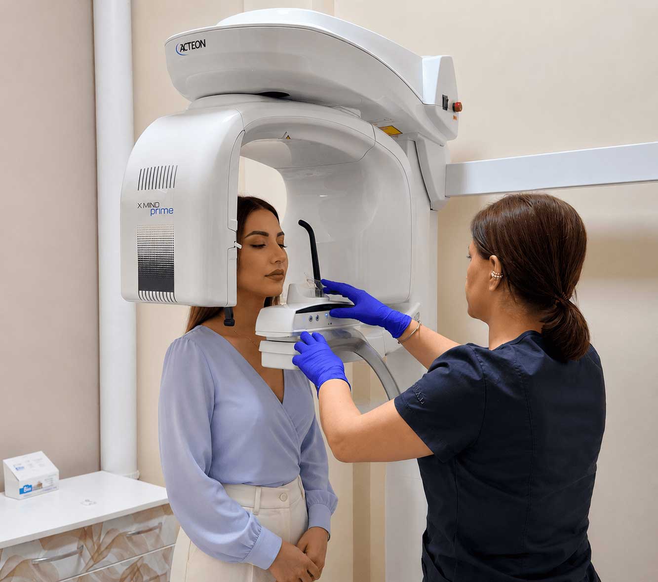
Cone-Beam Computed Tomography (CBCT) is an advanced diagnostic imaging technique that provides highly detailed three-dimensional (3D) images of the teeth, jaws, surrounding bone, and other oral and maxillofacial structures. It enables dental professionals to accurately evaluate the patient's anatomy, establish a precise diagnosis, and develop the most appropriate treatment plan.
During the examination, the CBCT scanner captures multiple images of the area of interest within just a few seconds. These images are then reconstructed into a detailed 3D model using specialized software, allowing the dentist to assess clinical conditions with exceptional accuracy and create a personalized treatment plan tailored to each patient's needs.
When is Maxillofacial Computed Tomography (CT) Necessary?
Maxillofacial computed tomography is prescribed in cases where obtaining more detailed information is essential for accurate treatment planning.
The dentist may recommend a CT scan for:
- Dental implant placement
- Wisdom tooth extraction
- Orthodontic treatment
- Surgical intervention of the jaws
- Detection of cysts or inflammatory focal points
- Evaluation of bone tissue volume
- Diagnosis of injuries or fractures
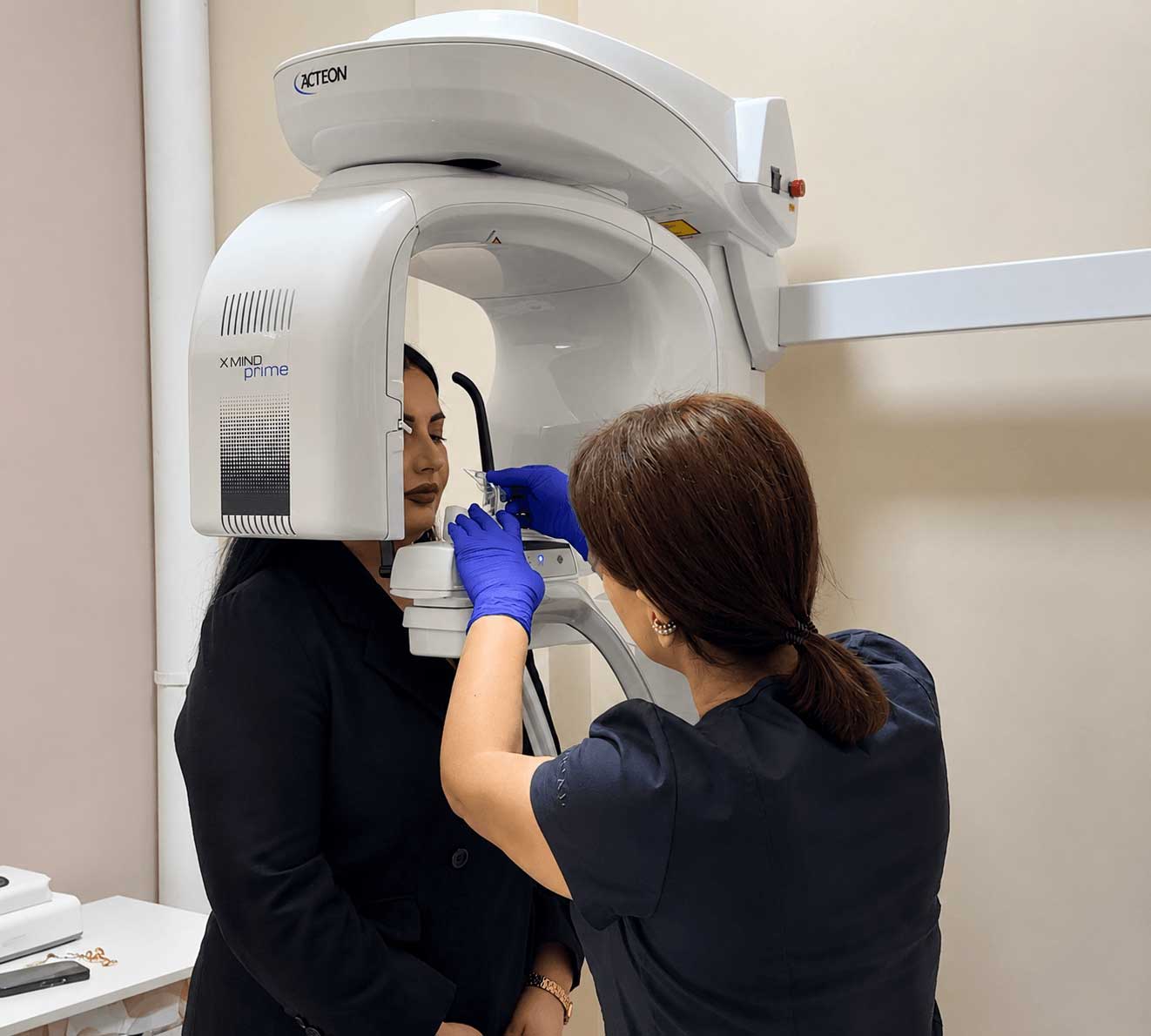
The examination is quick, comfortable, and non-invasive. Most scans take only a few seconds to complete while the scanner captures multiple images from different angles. Advanced software then reconstructs these images into a highly detailed 3D model, allowing your dentist to thoroughly evaluate the treatment area and develop the most appropriate treatment plan.
The Difference Between Dental X-rays and CT Scans
Both dental X-rays and Cone-Beam Computed Tomography (CBCT) play an essential role in modern dental diagnostics, but they serve different purposes and provide different levels of detail.
Conventional dental X-rays produce two-dimensional (2D) images, which are effective for routine examinations. However, because anatomical structures overlap in a 2D image, certain conditions—such as hidden infections, bone loss, impacted teeth, or the exact position of tooth roots—may not be fully visible.
In contrast, Cone-Beam Computed Tomography (CBCT) captures multiple images from different angles as the scanner rotates around the patient's head. These images are reconstructed into a highly detailed three-dimensional (3D) model, allowing the dentist to accurately evaluate the teeth, jaws, bone structures, nerve pathways, and surrounding anatomical features.
Key advantages of Dental CBCT include:
- High-resolution 3D imaging
- More detailed assessment of bone quality and anatomy
- Precise visualization of nerves and surrounding structures
- Improved planning for dental implants and oral surgery
- Greater diagnostic accuracy in complex clinical cases
While Cone-Beam Computed Tomography (CBCT) provides significantly more diagnostic information than conventional dental X-rays, it is not intended to replace them in every situation. The most appropriate imaging method is selected by the dentist based on each patient's individual clinical needs and treatment goals.
Dental CT Scan in Yerevan
If you are searching for a Dental cone beam CT scan near me, it is important to choose a clinic equipped with advanced imaging technology and experienced dental professionals.
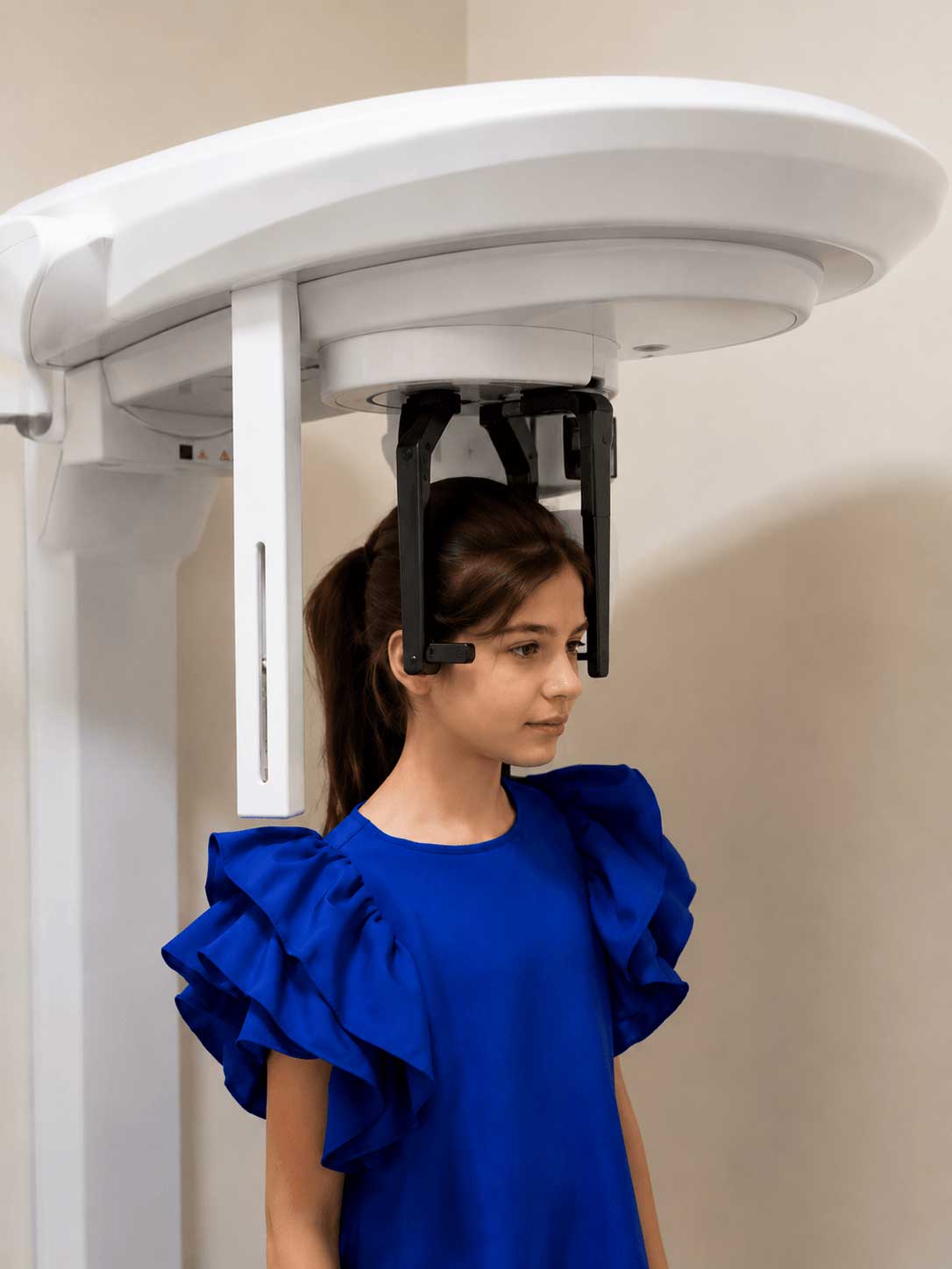
At MaryClinic, Cone-Beam Computed Tomography (CBCT) is performed using modern, high-precision equipment that delivers detailed three-dimensional (3D) images for accurate diagnosis and treatment planning. The examination is fast, comfortable, and completely non-invasive.
The cost of a CBCT scan depends on the area being examined. At MaryClinic, the price ranges from $24 to $55 USD, depending on whether the scan includes a single region, one jaw, or the entire maxillofacial area.
Why Choose MaryClinic?
When it comes to your oral health and smile, choosing the right dental clinic is just as important as choosing the right treatment.
At MaryClinic, we are convinced that accurate diagnosis is the foundation of successful treatment. For this reason, dental computed tomography (CT) is performed using state-of-the-art equipment from the Italian brand Acteon X-Mind, allowing us to obtain high-quality 3D images for precise treatment planning.
Every CBCT scan is carefully reviewed by our experienced dental professionals, allowing us to accurately assess each patient's clinical condition and recommend the most appropriate treatment approach. Whether you require dental implants, orthodontic treatment, oral surgery, or other specialized dental procedures, our team is committed to providing exceptional care at every stage of your treatment.
Our mission is to combine advanced technology, evidence-based dentistry, and a patient-centered approach to deliver safe, effective, and long-lasting results.
At MaryClinic, we are convinced that accurate diagnosis is the foundation of successful treatment. For this reason, dental computed tomography (CT) is performed using state-of-the-art equipment from the Italian brand Acteon X-Mind, allowing us to obtain high-quality 3D images for precise treatment planning.
Every CBCT scan is carefully reviewed by our experienced dental professionals, allowing us to accurately assess each patient's clinical condition and recommend the most appropriate treatment approach. Whether you require dental implants, orthodontic treatment, oral surgery, or other specialized dental procedures, our team is committed to providing exceptional care at every stage of your treatment.
Our mission is to combine advanced technology, evidence-based dentistry, and a patient-centered approach to deliver safe, effective, and long-lasting results.
Schedule your consultation at MaryClinic today and take the first step toward a healthier, more confident smile.


Frequently Asked Questions
-
"CT" stands for Computed Tomography. It is a 3D (three-dimensional) X-ray examination that allows the doctor to view a 3D image of your jaws and tooth roots, as well as their anatomical structure and bone tissue, from any angle and layer. Unlike a conventional 2D X-ray, a 3D CT scan shows a complete and flawless image without shadows or distortions.
-
It is completely safe and poses no risk to your health. Modern dental CT machines operate with minimal radiation exposure. For instance, the radiation dose received during a single scan is approximately equal to the amount a person receives during a regular airplane flight of a few hours.
-
In Yerevan's leading clinics and diagnostic centers, the cost of a 3D dental computed tomography scan ranges between 9,000 and 20,000 AMD, depending on which section of the jaw or jaws it covers.